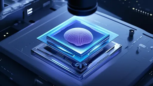
For decades, neuroscientists have grappled with the challenge of observing neurotransmitter activity in real time within intact biological systems. The dopaminergic pathway, a critical player in reward, motivation, and motor control, has remained particularly elusive due to technical limitations in tracking fast, microscopic signaling events deep within living tissue. A groundbreaking approach merging quantum nanotechnology with functional imaging – dubbed "Quantum Dot Neurocinema" – is now rewriting the rules of neurobiological observation.
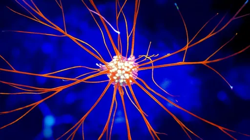
At the heart of this revolution are engineered semiconductor nanocrystals that fluoresce with exceptional brightness and photostability. Unlike traditional organic dyes that bleach within seconds or genetic calcium indicators with limited temporal resolution, these quantum dots can be targeted to specific neuronal populations and synaptic regions. When conjugated to dopamine-specific ligands or expressed as fusion proteins with dopamine receptors, they create an optical readout of neurotransmitter release and receptor activation across entire neural circuits.
The true breakthrough lies in the system's ability to operate in awake, behaving mammals. Early experiments in rodent models have produced stunning video-rate datasets showing dopamine waves propagating through the mesolimbic pathway during reward tasks. Researchers at the Max Planck Institute for Neuroscience recently captured never-before-seen patterns of striatal dopamine "microsurges" preceding lever presses in operant conditioning paradigms. These 50-100 millisecond events, invisible to conventional fast-scan cyclic voltammetry, suggest a finer temporal control of motivated behavior than previously imagined.
What makes the quantum dot approach transformative is its multiplexing capacity. By tuning different quantum dots to distinct emission wavelengths, teams at Stanford have simultaneously tracked dopamine, glutamate, and GABA transmission in the prefrontal cortex during decision-making tasks. This multi-neurotransmitter imaging reveals how dopaminergic signals are integrated with other systems at the synaptic level, providing the first direct evidence of "chemical computation" in cortical microcircuits.
Clinical applications are already emerging. A collaboration between MIT and Massachusetts General Hospital has adapted the technology for intraoperative use in Parkinson's disease patients undergoing deep brain stimulation. Quantum dot-labeled dopamine sensors injected into the subthalamic nucleus provide real-time feedback on stimulation efficacy, allowing neurosurgeons to optimize electrode placement based on neurotransmitter dynamics rather than just anatomical landmarks.
Nevertheless, significant challenges remain. The long-term biocompatibility of quantum materials requires further study, and current versions of the technology still lack the spatial resolution to visualize individual synaptic vesicles. Teams in Japan and Switzerland are reportedly developing next-generation "smart dots" that change both color and intensity based on dopamine concentration, potentially allowing absolute quantification of transmission events.
As the method spreads to primate labs and human clinical trials, it promises to reshape our understanding of addiction, depression, and neurodegenerative disorders. The ability to film neurotransmitter activity like a biological movie – complete with chemical close-ups and circuit-wide establishing shots – represents more than just a technical achievement. It offers neuroscientists what the telescope gave astronomers: a direct window into processes that were previously matters of inference rather than observation.
The coming years will likely see quantum dot imaging combined with other cutting-edge techniques. Early-stage work at Caltech has already paired it with optogenetics, allowing researchers to stimulate specific neuronal populations while watching the resulting dopamine release patterns. Other groups are working on miniaturized versions that could be integrated into wearable devices for chronic monitoring of neurotransmitter imbalances.
While still in its relative infancy, quantum dot neurocinema has already provided startling insights. Contrary to the classical view of dopamine as a slow, volume-transmitted modulator, the new data shows it operating with surprising speed and spatial precision. These observations are forcing a reconsideration of decades-old models about how reward signals are processed and propagated through the brain's networks.
From fundamental science to clinical neurology, the implications are profound. As the technology matures, we may soon have front-row seats to the biochemical ballet that underlies everything from creative inspiration to compulsive behavior – all rendered in vivid nanometer-scale color.

By /Aug 5, 2025
By /Aug 5, 2025

By /Aug 5, 2025

By /Aug 5, 2025

By /Aug 5, 2025

By /Aug 5, 2025

By /Aug 5, 2025

By /Aug 5, 2025

By /Aug 5, 2025

By /Aug 5, 2025

By /Aug 5, 2025
By /Aug 5, 2025
By /Aug 5, 2025
By /Aug 5, 2025

By /Aug 5, 2025

By /Aug 5, 2025

By /Aug 5, 2025

By /Aug 5, 2025

By /Aug 5, 2025

By /Aug 5, 2025